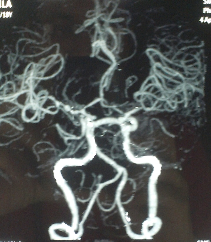
Moyamoya literally means “puff of smoke” in Japanese. It was named this because of its appearance on angiography, where it looks like a tangled network of new vessels in the brain. These new blood vessels appear adjacent to stenotic vessels. The steno-occlusive areas are usually bilateral but can occur unilaterally as well. 1
Moyamoya disease (or syndrome) is a progressive cerebrovascular disorder caused by blocked arteries at the base of the brain in the basal ganglia. It was first discovered in the 1960’s in Japan. Since then, it has been diagnosed in the US, Europe, Africa, and Australia. It most commonly affects children, but there have been cases found in adults as well. The presenting symptom is often a stroke or TIA, which are often accompanied with seizures and muscle weakness/paralysis on one side of the body. Adults tend to have hemorrhagic strokes. Other symptoms include visual disturbances, cognitive defects, abnormal consciousness, speech problems, sensory deficits and involuntary movements. It does appear to run in families so researchers suspect a genetic inheritance as a cause. 2
Moyamoya involves progressive narrowing of the carotid arteries at the base of the brain where they divide into the middle and anterior cerebral arteries. Specifically, the walls of the arteries thicken taking up space inside the lumen of the arteries and this narrowing can eventually result in complete blockage. To compensate for these narrowing arteries, new collateral vessels form to continue to supply oxygen-rich blood to the brain. These collaterals are much more fragile than normal vessels and break easier and can bleed into the brain. Aneurysms are often seen as well. “The progression of Moyamoya follows a typical course and can be classified into stages based on angiography findings.
- Stage I: Narrowing of internal carotid arteries
- Stage II: Development of moyamoya vessels at the base of the brain
- Stage III: Intensification of moyamoya vessels and internal carotid artery narrowing (most cases diagnosed at this stage)
- Stage IV: Minimization of moyamoya vessels and increased collateral vessels from the scalp
- Stage V: Reduction of moyamoya vessels and significant internal carotid artery narrowing
- Stage VI: Disappearance of moyamoya vessels, complete blockage of internal carotid arteries, and significant collateral vessels from the scalp “ 3
The prevalence of Moyamoya disease is <1 per 100,000. It tends to be highest in people of Asian descent, where it can reach as high as 3-4 per 100,000. The highest rate of occurrence is in the first decade of life and is slightly more common (1.8:1) in females. Death from this disease usually occurs because of hemorrhage. In childhood, there is a 4.3% mortality rate compared to adults where the mortality rate is 10%. Approximately 50-60% of patients experience progressive cognitive decline. 4
The diagnosis is established by typical findings on angiogram. CT scan or MRI may raise certain clues but it cannot be diagnosed definitively without an angiogram. The angiogram will show blockage in the internal carotid artery plus the “puff of smoke”. Additionally, PET scanning can reveal the areas of the brain with diminished blood supply. 5
Patients are often placed on aspirin to prevent stroke. However, the gold standard of treatment is surgical. Revascularization is done either directly or indirectly. In the direct revascularization approach (STA-MCA bypass) , a branch of a scalp artery is directly anastmosized to a branch of the middle cerebral artery (MCA) on the outside of the brain. This provides immediate improvement to blood supply to the brain. An indirect revascularization is achieved as well by lying on the outside of the brain. The improvement in blood flow is expected to continue improving over several months. Results at the Moyamoya center have shown a 95 % graft patency rate with good long-term clinical outcomes. There are various indirect revascularization methods using different arteries and even using the temporalis muscle. The results are mixed and take longer to achieve. 6
There are several research studies being conducted currently about this disease.
- 1 http://emedicine.medscape.com/article/1180952-overvie
- 2 http://www.ninds.nih.gov/disorders/moyamoya/moyamoya.htm
- 3 http://www.mayfieldclinic.com/PE-Moyamoya.htm#.VgLPZd9Viko
- 4 http://emedicine.medscape.com/article/1180952-overview#a7
- 5 http://www.columbianeurosurgery.org/conditions/moyamoya-disease/
- 6 http://neurosurgery.stanford.edu/moyamoya/treatments.html
ORIGINALLY POSTER ON SERMO
 Copyright secured by Digiprove © 2016 Linda Girgis, MD, FAAFP


